Home
Home
How 600 Human Muscles Orchestrate Every Movement From Smiling to Sprinting
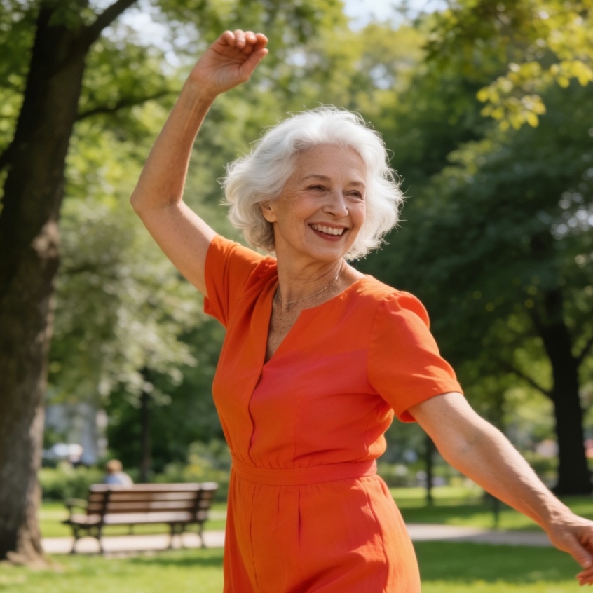
The human body functions as a complex biological masterpiece, and at the center of its mechanical prowess is the muscular system. Comprised of more than 600 individual muscles, this network is responsible for everything from the subconscious beating of the heart to the explosive power required for a world-class sprint. While the skeleton provides the structural framework, the muscles are the engines that pull, stabilize, and protect.
Understanding the muscular system requires looking beyond simple movement. Muscles are not merely meat and fiber; they are sophisticated transducers that convert chemical energy from food into mechanical work. In an average adult, muscle mass accounts for approximately 30% to 40% of total body weight, serving as the body’s primary consumer of oxygen and nutrients.
The Three Biological Architectures of Human Muscle
To understand how the body moves, one must first distinguish between the three primary types of muscle tissue. Each has a distinct cellular structure and a unique role within the human physiological economy.
Skeletal Muscle and Voluntary Control
Skeletal muscles are what most people refer to when they talk about "muscles." These are the organs of the muscular system that attach to bones via tendons. Their primary characteristic is that they are under voluntary control. When you decide to lift a coffee mug or kick a ball, your brain sends an electrical impulse through the nervous system to specific skeletal muscles.
Under a microscope, skeletal muscle appears striated or striped. This is due to the highly organized arrangement of actin and myosin filaments, which are the proteins responsible for contraction. Because these muscles often cross joints, their contraction results in the movement of the skeleton. In our practical observations of human movement, skeletal muscles are unique because they can be trained, hypertrophied through exercise, and fatigued through repetitive use.
Cardiac Muscle and the Infinite Pulse
Cardiac muscle is found exclusively in the heart. Like skeletal muscle, it is striated, but it operates under involuntary control. You do not have to "think" about making your heart beat; the sinoatrial node—the heart's natural pacemaker—regulates this rhythm automatically.
The most fascinating aspect of cardiac muscle is its resistance to fatigue. While a leg muscle might cramp after an hour of running, cardiac muscle cells are packed with mitochondria, the energy-producing powerhouses of the cell, allowing the heart to contract roughly 100,000 times a day for a lifetime without rest.
Smooth Muscle and the Silent Operators
Smooth muscle is located in the walls of hollow organs, such as the stomach, intestines, and blood vessels. Unlike the other two types, it lacks striations, giving it a "smooth" appearance. These muscles are involuntary and are controlled by the autonomic nervous system.
Smooth muscle performs the quiet, essential tasks of survival. It moves food through the digestive tract via a process called peristalsis and regulates blood pressure by contracting or dilating blood vessels. While we rarely feel smooth muscles working, their failure—such as during a vascular spasm or an intestinal cramp—can cause significant physical distress.
Decoding the Names of the Muscular System
The nomenclature of human muscles can seem like a foreign language, but it follows a highly logical system. Most muscle names provide a "GPS coordinate" or a functional description of the tissue. Understanding these naming conventions makes anatomy far more intuitive.
- By Location: The frontalis muscle is located on the frontal bone of the skull. The tibialis anterior is found on the front of the tibia (shin bone).
- By Size: Terms like maximus (largest), minimus (smallest), longus (long), and brevis (short) are common. For example, the gluteus maximus is the largest muscle in the buttock region.
- By Shape: The deltoid is named after the Greek letter delta because of its triangular shape. The trapezius is shaped like a trapezoid.
- By Direction of Fibers: Rectus means straight; thus, the rectus abdominis has fibers that run vertically. Oblique refers to fibers at an angle.
- By Number of Origins: Biceps means "two heads," triceps means "three heads," and quadriceps means "four heads." These names tell you exactly how many attachment points the muscle has at its origin.
- By Action: The adductor longus moves the leg toward the midline of the body (adduction), while the extensor digitorum extends the fingers.
The Mechanics of Movement and Muscle Pairing
Muscles are incapable of "pushing." They can only contract (shorten) and relax (lengthen). To move a bone back and forth, muscles must work in antagonistic pairs.
Agonists and Antagonists
The muscle that is primarily responsible for a movement is called the agonist or prime mover. The muscle that opposes that movement is the antagonist. In our practical observation of an arm curl, the biceps brachii acts as the agonist, contracting to bend the elbow. Simultaneously, the triceps brachii on the back of the arm must relax and elongate to allow the movement to happen. If you were to extend your arm back to a straight position, the roles would reverse: the triceps would become the agonist and the biceps the antagonist.
Synergists and Fixators
Movement is rarely a solo act. Synergists are muscles that assist the prime mover. For instance, when the biceps flexes the elbow, the brachialis and brachioradialis provide additional force. Fixators are specialized synergists that stabilize the origin of the prime mover. When you lift a heavy weight, the muscles around your shoulder and scapula act as fixators, holding the bone steady so the arm muscles have a firm base from which to pull.
A Regional Tour of the Body Major Muscle Groups
To appreciate the scale of the muscular system, we must look at how these tissues are distributed across the body’s different regions.
The Face and Neck: The Hardware of Expression
The muscles of the head are unique because many of them attach to the skin rather than to bone, allowing for the vast range of human facial expressions.
- Frontalis: This muscle allows you to raise your eyebrows and wrinkle your forehead in surprise.
- Orbicularis Oculi: A circular muscle that surrounds the eye, enabling you to blink, squint, or close your eyes tightly.
- Zygomaticus: Often called the "smiling muscle," it pulls the corners of the mouth upward.
- Masseter: Located in the jaw, this is technically the strongest muscle in the human body based on its weight. It provides the incredible force needed for chewing.
- Sternocleidomastoid: A major muscle of the neck that allows you to rotate your head from side to side or tilt it forward.
The Core and Trunk: The Stability Center
The trunk muscles protect the internal organs and maintain the upright posture that defines the human species.
- Pectoralis Major: The large, fan-shaped muscle of the chest used for pushing movements and pulling the arms toward the center of the body.
- Rectus Abdominis: Often called the "six-pack," these muscles flex the lumbar spine. During our physical assessments, we observe that these are critical for core stability and intra-abdominal pressure.
- External and Internal Obliques: Located on the sides of the abdomen, these allow for twisting the torso and side-bending.
- Latissimus Dorsi: The largest muscle of the upper body, the "lats" are responsible for pulling movements, such as those used in swimming or pulling a door open.
- Trapezius: A large diamond-shaped muscle on the back that moves the shoulder blades and supports the neck.
Upper Extremities: Precision and Power
The muscles of the arms and hands are designed for a combination of heavy lifting and fine motor skills.
- Deltoid: The thick muscle that gives the shoulder its rounded shape. It is the primary mover for lifting the arm out to the side.
- Biceps Brachii and Triceps Brachii: The famous pair that controls the flexion and extension of the elbow.
- Rotator Cuff: A group of four muscles (supraspinatus, infraspinatus, teres minor, and subscapularis) that stabilize the highly mobile shoulder joint. In clinical experience, these are among the most common sites for sports-related injuries.
- Forearm Flexors and Extensors: These muscles control the complex movements of the wrist and fingers. The human hand contains more than 30 muscles (some originating in the forearm), allowing for the "precision grip" that enabled humans to develop tools.
Lower Extremities: The Foundation of Locomotion
The muscles of the legs are the largest and most powerful in the body, designed to move our entire body weight through space.
- Gluteus Maximus: This muscle is essential for maintaining an upright posture and providing the power to climb stairs or run. It is one of the thickest muscles in the body.
- Quadriceps Femoris: A group of four muscles on the front of the thigh. They are the primary extensors of the knee. In a practical gym setting, you feel these muscles "burn" during squats or lunges.
- Hamstrings: A group of three muscles on the back of the thigh that flex the knee and extend the hip.
- Gastrocnemius and Soleus: These form the calf. The gastrocnemius gives the calf its shape and provides the power to push off the ground during walking, while the deeper soleus is vital for standing balance.
- Sartorius: The longest muscle in the human body, it runs diagonally across the thigh and helps with both hip and knee flexion.
The Science of Muscle Contraction and Energy
At the microscopic level, muscle contraction is an elegant chemical dance. This process is described by the Sliding Filament Theory. Inside each muscle fiber are thousands of smaller structures called myofibrils, which contain segments called sarcomeres.
When a nerve signal reaches the muscle, it triggers the release of calcium ions. These ions allow the protein heads of myosin to "grab" onto the actin filaments and pull them inward. This shortening of the sarcomere is what we perceive as a muscle contraction.
This process requires a constant supply of ATP (Adenosine Triphosphate). To produce ATP, muscles utilize different energy systems:
- Phosphagen System: For short, explosive bursts of power (0-10 seconds).
- Glycolytic System: For high-intensity efforts lasting up to two minutes (often resulting in the "burn" of lactic acid).
- Aerobic System: For long-duration, lower-intensity activities like walking or distance running, utilizing oxygen to burn fats and carbohydrates.
Essential Functions Beyond Simple Movement
While locomotion is the most obvious role of the muscular system, it performs several "invisible" functions that are vital for survival.
Heat Production (Thermogenesis)
Muscles are inefficient engines; when they contract, a large portion of the energy is lost as heat. This is actually a biological advantage. When you are cold, your brain triggers involuntary muscle contractions known as shivering to generate heat and maintain your core body temperature.
Posture and Stability
Even when you are standing perfectly still, your muscles are hard at work. Small "tonic" contractions in the back, neck, and legs prevent gravity from pulling you to the floor. This constant tension is what allows us to maintain an upright posture for hours at a time.
Protection and Support
Abdominal muscles, for example, have no bone behind them to protect the intestines and liver. Instead, layers of muscle arranged in different fiber directions create a "biological corset" that protects internal organs from impact and keeps them in their proper anatomical positions.
Maintaining Muscular Health for Long-Term Mobility
Based on our observations of aging and physical performance, muscle mass naturally begins to decline after the age of 30—a process known as sarcopenia. However, this is not inevitable.
- Resistance Training: Placing muscles under tension through weightlifting or bodyweight exercises stimulates muscle protein synthesis, maintaining strength and bone density.
- Protein Intake: Muscles require amino acids to repair the micro-tears caused by exercise. A diet rich in high-quality protein is essential for recovery.
- Flexibility and Mobility: Muscles that are never stretched can become "short" and tight, leading to joint pain and reduced range of motion.
- Hydration and Electrolytes: Muscles require water, sodium, potassium, and magnesium to conduct electrical impulses. Dehydration is the most frequent cause of muscle cramps in athletes.
Summary of the Human Muscular System
The human muscular system is a vast network of over 600 muscles classified into skeletal, cardiac, and smooth tissues. While skeletal muscles provide voluntary movement through antagonistic pairing, cardiac and smooth muscles manage the involuntary processes of circulation and digestion. Each muscle is named based on logical criteria such as size, location, or function, and they all work together to provide stability, generate heat, and enable the staggering variety of human activities. From the tiny stapedius muscle in the ear to the massive gluteus maximus, every fiber plays a role in the symphony of human life.
Frequently Asked Questions about Human Muscles
How many muscles are in the human body?
Most medical texts state there are between 600 and 650 muscles. The exact number can vary depending on whether certain muscle groups are counted as a single unit or as individual parts.
What is the strongest muscle in the body?
There is no single answer, as "strength" can be measured in different ways. The masseter (jaw muscle) is the strongest based on weight-to-force ratio. The gluteus maximus is the largest and provides the most absolute force. The heart is the strongest in terms of endurance and continuous work.
What is the smallest muscle in the body?
The stapedius is the smallest muscle. Located in the middle ear, it is only about 1.27 millimeters long. Its function is to stabilize the smallest bone in the body, the stapes, to protect the ear from loud noises.
Why do muscles get sore a day or two after exercise?
This is known as Delayed Onset Muscle Soreness (DOMS). It is caused by microscopic tears in the muscle fibers and the subsequent inflammatory response as the body repairs and strengthens the tissue. It is not caused by lactic acid, which usually leaves the muscle within an hour after exercise.
Can muscles turn into fat if you stop exercising?
No. Muscle and fat are two entirely different types of tissue. If you stop exercising and maintain a caloric surplus, muscle cells will shrink (atrophy) and fat cells will grow (hypertrophy), but one tissue never "turns into" the other.
-
Topic: Muscles of the Human Body | Anatomy, Diagram & Functions - Lesson | Study.comhttps://study.com/academy/lesson/major-muscle-groups-of-the-human-body.html
-
Topic: Muscular System | Anatomy Gallery | American Cancer Societyhttps://www.cancer.org/cancer/understanding-cancer/anatomy-gallery/muscular-system.html
-
Topic: 9: MUSCULAR SYSTEMhttps://batch.libretexts.org/print/Finished/med-63433/Full.pdf